Correlation between GATA3 Immunohitsochemistry and TP53 on some Ovarium Carcinoma Histopathology Subtype
DOI:
https://doi.org/10.55816/mpi.v34i3.665Keywords:
Clear cell carcinoma, Endometrioid carcinoma, GATA3, karsinoma ovarium, Mucinous carcinoma, Serous carcinoma, p53Abstract
Background
Ovarian carcinoma is a cancer with high mortality in women, although comprehensive treatment with surgery and chemotherapy is at an advanced stage, survival rates are still low. GATA3 and p53 are predictors of some malignancies, but results vary in ovarian carcinoma.
Objective
To examine correlation between immunohistochemical expression of GATA3 and p53 in patients with ovarian carcinoma with various histopathological subtypes.
Materials and Methods
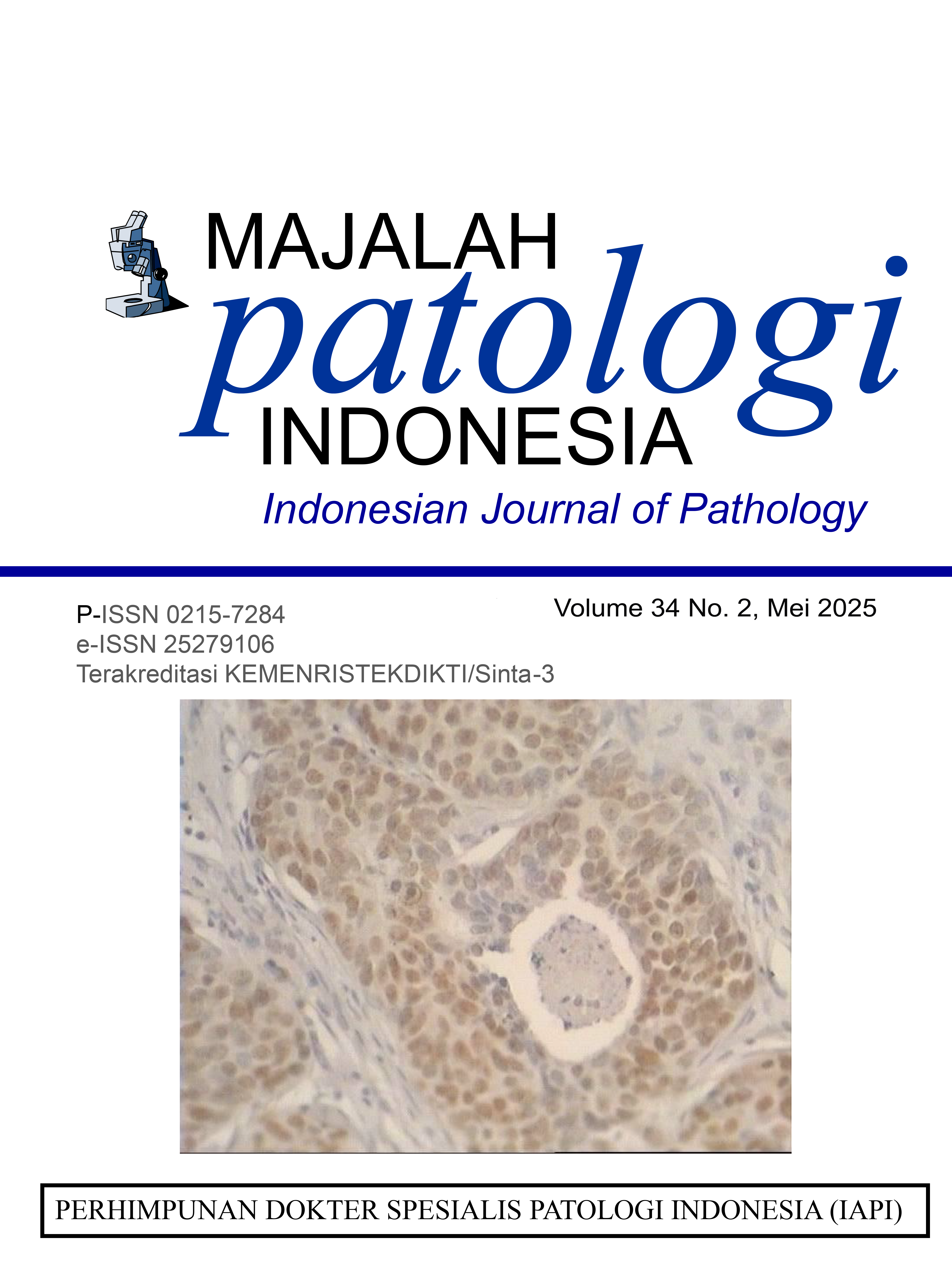
Cross sectional design is the method of this study conducted on slides of 28 ovarian carcinoma patients in several subtypes of hitopathology. Each slide was stained with hematoxylin and eosin (H&E) to assesses hitopathological subtypes and stained with GATA3 and p53 antibodies. Expression GATA3 was assessed using H-score and p53 quick score. A logistic regression assay (p<0.005) was used to assessed the association of GATA3 and TP53 immunohistochemical expression in several histopathological subtypes of ovarian carcinoma. Statistical analysis between GATA3 and p53 was performed using the eta correlation test is used because the data is nominal-ordinal.
Results
Among 28 specimens in patients with ovarian carcinoma, Cases was most prevalent in the age group >50-60 years (age range 58 years), history of nullipara parity, and most in the group of stage III ovarian malignancy. Positive immunohistochemical p53 expression is more prevalent in serous carcinoma. Positive GATA3 immunohistochemical expression is more prevalent in serous carcinoma.
Conclusion
There is no significant relationship. Immunohistochemical expression of GATA3 and TP53 in some histopathological subtypes of ovarian carcinoma. However, immunohistochemical expression of GATA3 high p53 positive tends to be found in high-grade serous carcinoma.
Keywodrs: GATA3, p53, karsinoma ovarium, Serous carcinoma, Mucinous carcinoma, Endometrioid carcinoma, Clear cell carcinoma.
Downloads
Downloads
Published
Issue
Section
License
Copyright (c) 2025 intan nefia alamanda, Betty Betty, Delyuzar Delyuzar, Jessy Chrestella, Soekimin Soekimin

This work is licensed under a Creative Commons Attribution-NonCommercial-NoDerivatives 4.0 International License.


.jpg)