Comparison of Peritumoral Budding Features in Colorectal Adenocarcinoma NOS by Hematoxylin-Eosin (HE) Staining and Pancytokeratin (AE1/AE3) Immunohistochemistry
DOI:
https://doi.org/10.55816/mpi.v34i2.639Keywords:
Colorectal adenocarcinoma NOS, peritumoral budding, HE, AE1/AE3Abstract
Background
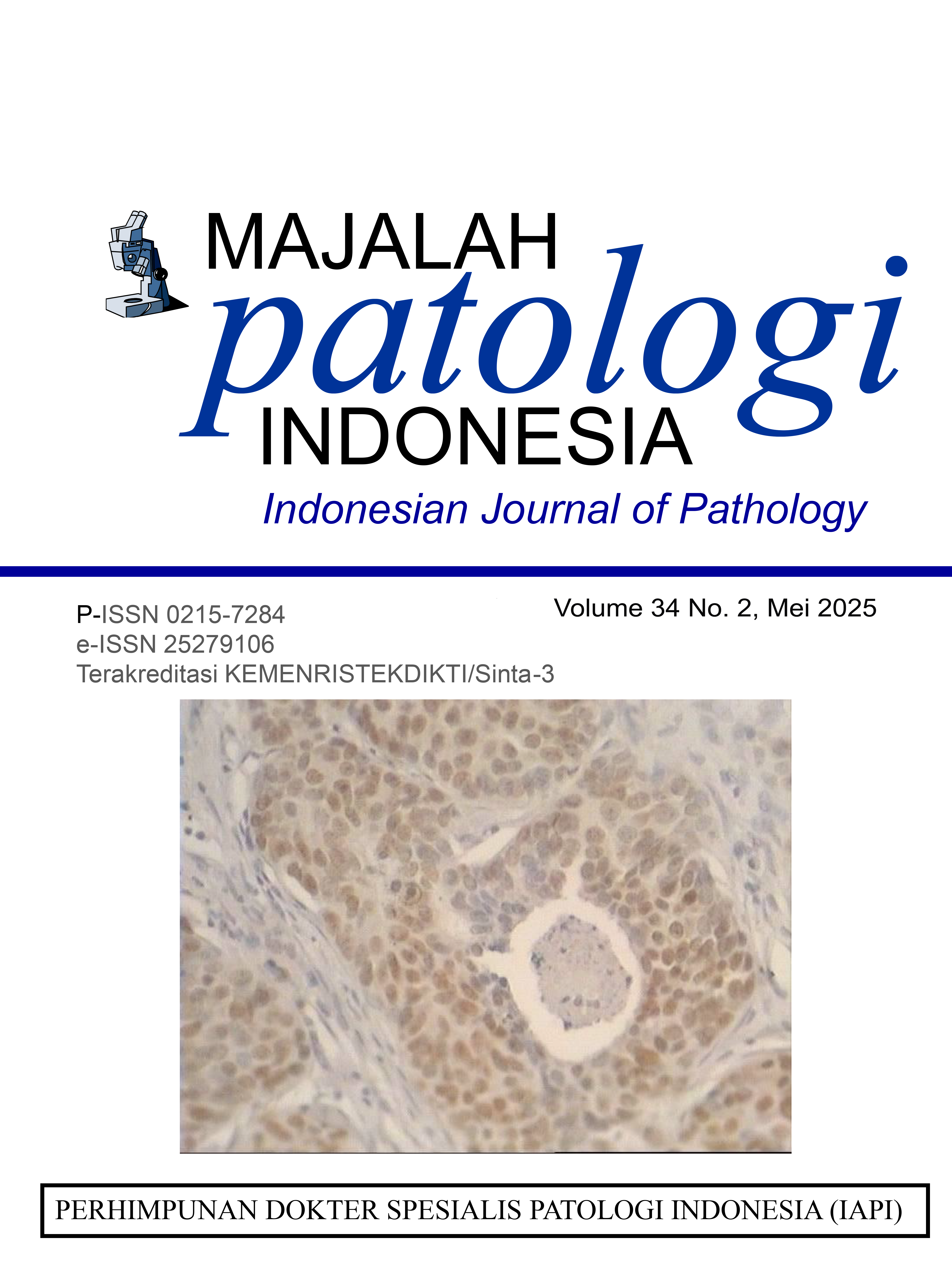
Colorectal adenocarcinoma is defined as a malignant epithelial tumor of the large intestine (colon and rectum) that shows glandular and mucinous differentiation, accompanied by invasion through the muscular mucosae into the submucosal layer. Peritumoral budding refers to tumor budding at the leading edge of the tumor and can be considered one of the prognostic factors. Immunohistochemistry Pancytokeratin (AE1/AE3) is observed in the epithelium, and most carcinomas (tumors originating from epithelial cells) are stained in the cytoplasm.
Method
The analytical study involved 48 paraffin block samples diagnosed as colorectal adenocarcinoma NOS at Haji Adam Malik Central General Hospital in Medan and the laboratory of the Faculty of Medicine, Universitas Sumatera Utara. The assessment of tumor budding using hematoxylin-eosin staining and pan-cytokeratin (AE1/AE3) immunohistochemical staining was classified equally into three categories: low budding category if 0-4 buds of tumor budding were observed, intermediate budding category if 5-9 buds of tumor budding were observed, and high budding category if ≥10 buds of tumor budding were observed.
Results
There is no difference in assessing peritumoral budding using Hematoxylin-Eosin (HE) staining and pan-cytokeratin (AE1/AE3) immunohistochemical staining.
Conclusion
Assessment of peritumoral budding is recommended using Hematoxylin-Eosin (HE) staining.
Downloads
Downloads
Published
Issue
Section
License
Copyright (c) 2025 listiyaningsih listiyaningsih

This work is licensed under a Creative Commons Attribution-NonCommercial-NoDerivatives 4.0 International License.


.jpg)